


🧬 Topic: Developmental biology · How the head, hindbrain and trunk are put together
🧩 The Modular Mystery of the Vertebrate Body
Have you ever wondered how the intricate bodies of vertebrates—whether they're fish, birds, mice, or humans—manage to create their repeating structural patterns from head to tail? This fascinating phenomenon, known as metamerism, is evident in our segmented skeletons, muscles, blood vessels, and nervous systems. For more than a century, scientists have grappled with the challenge of understanding how this architectural design unfolds during embryonic development.
In the past, comparative anatomists like E.S. Goodrich and H.C. Bjerring proposed that vertebrates are fundamentally segmented in a consistent manner from the tip of the head all the way to the tail. They believed that the segmentation in the head was simply obscured over time due to the specialized needs of cephalization and the process of embryonic development.
🔎 The big shift
Recent advances in molecular biology have shaken up this long-held belief in a single, uniform blueprint. By delving into the periodic expression of regulatory genes, tracing cell lineages, and setting strict criteria for metamerism, researchers have uncovered that the vertebrate body axis is actually a modular patchwork. Instead of following one grand design, evolution employs a variety of region-specific molecular tricks to construct the head and trunk.
🧠 Your head isn't just a simple extension of your spine—it's more like an evolutionary "add-on."
Traditionally, anatomy textbooks depicted the head as a specialized continuation of the segmented trunk. However, a groundbreaking evolutionary model from Carl Gans and R. Glenn Northcutt paints a different picture, one that modern cell lineage tracing supports.
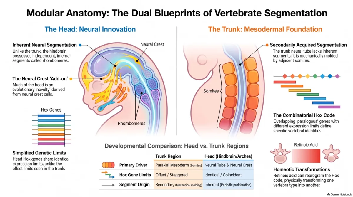
Researchers pointed out that in early chordates like Amphioxus, the notochord stretches all the way to the very front of the organism. In contrast, in vertebrates, the notochord ends much further back, around the mid-brain area. The front part of the vertebrate head is mainly made up of tissues that come from developmental innovations: the neural crest and cranial placodes. Since these structures emerged alongside new feeding mechanisms during the early evolution of vertebrates, Gans and Northcutt concluded that the majority of the front head is actually unsegmented.
Shifting from the traditional view of a seamless head-to-tail connection to a model of modular "add-ons" significantly alters our understanding of evolutionary history. It reveals that the front of the head isn't just a series of modified spinal segments; rather, it's a completely new anatomical area added onto an ancient chordate body plan. 🦴
🧪 A Single Chemical Can Rewrite Your Skeletal Blueprint
In the trunk mesoderm, the identity of different regions along the vertebral column is determined by a fascinating system known as the "Hox code." This code consists of Antennapedia class homeobox genes that are organized in clusters on chromosomes, and their expression plays a crucial role in defining the shape and identity of various anatomical regions, like cervical, thoracic, and lumbar areas.
So, what happens when this regulatory barcode gets disrupted? Researchers Michael Kessel and Peter Gruss showcased the incredible influence of this system by using retinoic acid, a potent morphogen. When they exposed mouse embryos to retinoic acid at specific stages of development, they found that it systematically changed the spatial expression patterns of the Hox genes.
Importantly, this chemical exposure didn't lead to chaotic malformations. Instead, it triggered precise homeotic transformations, reshaping entire anatomical segments into alternative, fully developed structures. For example, by altering the Hox code, lumbar vertebrae were transformed into rib-bearing thoracic structures, increasing the number of thoracic vertebrae in treated mice from 13 to 15.
13 → 15 THORACIC VERTEBRAE IN TREATED MICE |
Retinoic acid THE MORPHOGEN USED |
|---|
The ability of a single chemical agent to systematically rewrite skeletal architecture underscores the modular nature of development. Minor adjustments in regulatory signaling don't result in random structural failures; rather, they effectively flip developmental switches, prompting embryonic cells to follow alternative, complete, and pre-programmed anatomical blueprints. 🎛️
🏗️ Reimagining the Architecture of Life
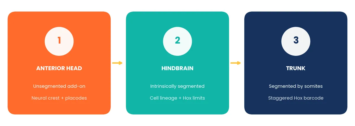
The vertebrate body axis isn't just a straightforward head-to-tail assembly; it's much more complex. Recent molecular findings show that our structure is actually a tri-partite system made up of at least three fundamentally different domains:
The Anterior Head: This part is an unsegmented, evolutionary "add-on," primarily formed from neural crest and placodal innovations.
The Hindbrain: Here, we find an intrinsically segmented neural domain, organized by strict cell lineage boundaries and specific Hox expression limits.
The Trunk: In this domain, secondary neural segmentation is shaped by mesodermal somites, influenced by staggered Hox genetic barcodes.
🌱 Food for thought
When we dig deeper into our anatomy, molecular biology uncovers that our bodies are built from a patchwork of unique historical innovations. Just think about it—how many other hidden treasures from our distant evolutionary past are still waiting to be discovered inside our biological system?
References
- Thorogood, Peter, and James Hanken. "Pattern Formation: Body Building Exercises." Current Biology, vol. 2, no. 2, 1992, pp. 83–85.